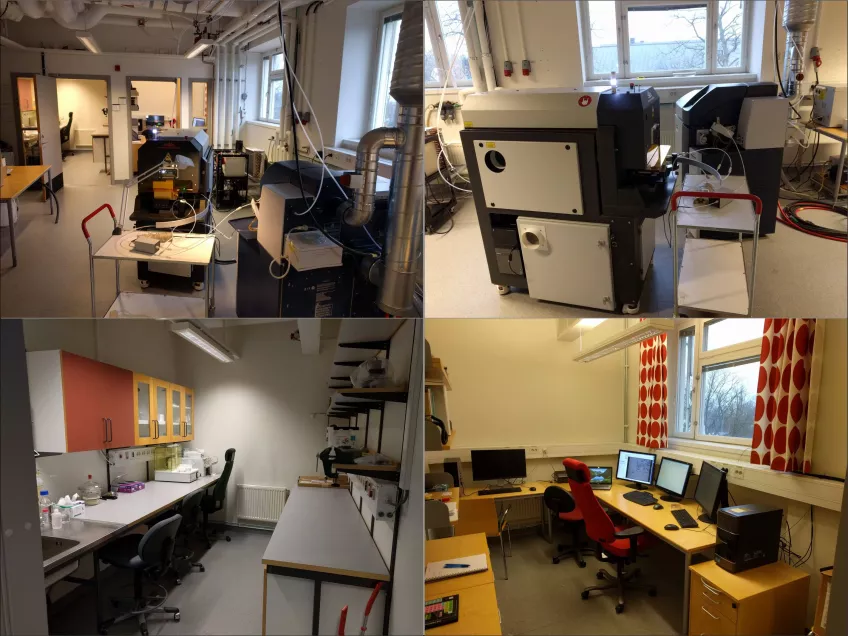
Laser ablation inductively coupled plasma mass spectrometry (LA-ICP-MS)
The laboratory is equipped with a Bruker Aurora Elite ICP-MS and a 193 nm Cetac Analyte G2 excimer laser installed with a two volume HelEx2 sample cell. LA-ICP-MS is a powerful method for element analysis in a wide variety of solid materials (i.e. minerals, bone & teeth materials, wood, ceramics, etc.) at spatial resolutions from a few up to several hundred µm. Quantitative analyses can be obtained for most elements in the mass range 5-250 amu (practically 7Li to 238U).
Sample introduction to the ICP-MS system is setup for both liquid samples by nebulizer and solid material by laser ablation.
The laboratory have a number of analytical procedures that are done routinely, which are listed below. We are always interested in developing new methods together with users of the laboratory, so please contact us to discuss your project. At the lab we use the software Iolite for data reduction.
Contact Tomas Næraa (tomas [dot] naeraa [at] geol [dot] lu [dot] se)
U-Pb isotope age dating
We have methods set up for precise U-Pb ages for a number of uranium bearing accessory minerals such as zircon, monazite, baddeleyite, rutile, titanite and apatite among others.
Routine methods at the lab
- Zircon U-Pb: We routinely use reference zircon GJ1 and 91500 as primary and secondary standards. Depending on zircon grain size and internal textures, spot size is routinely set between 20 and 35 µm.
- Monazite U-Pb: For this method we use monazite 44069 as primary standard and we are in the process of developing several in-house reference monazites. Spot sizes are in the range of ca. 5 to 15 µm.
- Titanite U-Pb: For this method we use MKED1, ONT2 and Khan as reference materials. Spot size range from 25 µm.
- Baddeleyite U-Pb: This method is under development. Our test results show that we can ablate with ca. 20 µm spot size and obtain sample average 207Pb/206Pb age precision around 0.3-0.5 %.
Trace element
Trace element analyses are possible for most solid materials; the main limiting factor is weather materials can be ablated with the laser beam. Another factor is the quantification of element concentrations; this can be done if matrix matched material of known concentration is available. However for many silicate materials, NIST standard glasses are sufficient for element quantification.
Routine methods
- Silicates, e.g. garnet, zircon and epidote, calibration using NIST 610-612-614 glass and USGS basaltic glasses (e.g. BCR2G/BHVO)
- Carbonate, calibration using MACS3, JCp-1 and JCt-1
- Titanite, calibration using MKED1, ONT2 and Khan natural titanite
- Chromite, calibration using inhouse reference material together with NIST glass
- Sulfide, calibration using MASS1.
Trace elemet maps
Trace element maps by LA-ICP-MS methods opens for relatively fast aquisation of spartial distribution of trace element data. Below example are from an otolith (fish ear-stone and a clino-pyroxene. Trace element mapping is an area under fast development and a focus area for this laboratory.
Profiles
Reference materials at the lab
- NIST glasses - 610, 612 and 614
- BCR - basalt glass
- BHVO - basalt glass
- MASS-1 - sulfide
- MACS-3 – carbonate
- JCp-1 - carbonate
- JCt-1 - carbonate
- GJ1 - natural zircon for U-Pb
- 91500 - natural zircon for U-Pb
- 44069 - natural monazite for U-Pb + several natural in-house materials
- MKED1 - natural titanite for U-Pb and trace elements
- Baddeleyite (several natural in-house reference samples)
Laser spot dimensions
The laser system has two different apertures for controlling beam size and shape. The mask aperture provides predefined circular and square spots with sizes that range from 1 to 155 µm; these spots can further be expanded through a lens which increases the size 1.6 times to a max of 258 µm. The x-y aperture provides similar sized spots but with a continous size range as squares or rectangles which can rotate 180 degrees.
Tubing and cones
In laser ablation, material is transported in EFTE tubing, a “squid” for signal smoothing are used for applications where low ablation rates are required. The lab is also equited with a ARIS (Aerosol Rapid Introduction System) by which it is possibly to resolve single pulses at sample rates up to 60 Hz, however this is not standard setup at our instrument. The lab operated with several sets of nickel cones that are used for different applications with different baseline requirements.
Sample holders and preparation
The laser lab have three different sample holders, one that holds 9 one inch mounts (2.54 cm in diameter), one that holds two to four thin-sections and three one inch mounts and one for odd size samples (10*10 cm and max 1 cm high).
In order to avoid mistakes and to obtain the best results possible, please consult the laboratory prior to your sample preparation.
The software that controls the laser can work on several image layers (e.g. images from SEM or light microscope) for easier navigation on samples. Also the software is setup for fast and easy imaging and mosaic imaging of samples. Proper image preparation will greatly help your work and optimize your analytical time at the instrument, and we are happy to help with the image preparation.


